Keynote speakers
Mackenzie Mathis

Scott Delp
Molly Stevens
ESB 2025 Keynote talks:
Towards the Neural Basis of Adaptive Motor Control
The neural activity of the brain is intimately coupled to the dynamics of the body. The motor system, which controls our outward behavior, spans multiple levels of hierarchical sensorimotor control: from the spinal cord, cerebellum, to the cortex and brainstem, at all levels, motor action is orchestrated. Yet we are experimentally limited to observing a small subset of all neurons of this hierarchy. Moreover, a mechanistic understanding of this complicated system is unknown, as mapping high-level behavior to neural activity is unconstrained. To address this, we built a large-scale model (MausSpaun) that captures hypothesized neural computations and coupled this with a novel model of the adult mouse forelimb in a physics simulation environment. We now use this model to better understand cortical computations during motor learning, which I will detail in my lecture.
Biography: Prof. Mackenzie W. Mathis is the Bertarelli Foundation Chair of Integrative Neuroscience and an Assistant Professor at the Swiss Federal Institute of Technology, Lausanne (EPFL). Following the award of her PhD at Harvard University in 2017 with Prof. Naoshige Uchida, she was awarded the prestigious Rowland Fellowship at Harvard to start her independent laboratory at Harvard (2017-2020). She is an ELLIS Scholar, Vallee Scholar, a former NSF Graduate Fellow, and her work has been featured in the news at Bloomberg BusinessWeek, Nature, and The Atlantic. She was awarded the FENS EJN Young Investigator Prize 2022 & the Eric Kandel Young Neuroscientist Prize in 2023. Her lab works on mechanisms underlying adaptive behavior in intelligent systems. Specifically, the laboratory combines machine learning, computer vision, and experimental work in rodents with the combined goal of understanding the neural basis of adaptive motor control.
Frontiers in Human PerformanceResearch: Insights from Biomechanical Simulation and Machine Learning
Rapid technical advancements in mobile sensing, computer vision, and data science have begun to enable planetary-scale experiments and major advancements in human health. In the next few years, these ongoing technical strides will culminate in a watershed moment for biomechanics and human performance research, when simulations run in real-time and AI becomes mainstream in studies of human movement and clinical applications. During my lecture, I will illustrate how our expanded capacity to amass large-scale datasets that characterize human movement, coupled with our accelerated ability to derive insights from automated data analyses, will inform clinical decisions and facilitate personalized training.
Biography: Scott Delp is the James H. Clark Professor of Bioengineering, Mechanical Engineering, and Orthopaedic Surgery at Stanford University. He is the Founding Chairman of the Department of Bioengineering at Stanford and Director of the Wu Tsai Human Performance Alliance, which aims to transform human health through the science of peak performance. Dr. Delp is also the Director of the RESTORE Center, a NIH national center focused on measuring real world rehabilitation outcomes and Director of the Mobilize Center, a NIH National Center of Excellence focused on Big Data and Digital Health. Scott’s laboratory develops technologies to advance movement science and human health. Software tools created in his lab, including OpenSim, OpenCap, AddBiomechanics, and Simtk.org, have become the basis of an international collaboration involving thousands of scientists. He has published over 300 research articles and has recently released a book from MIT Press entitled Biomechanics of Movement: The Science of Sports, Robotics, and Rehabilitation. Dr. Delp has co-founded six health technology companies and is a member of the U.S. National Academy of Engineering.
Designing Biomaterials for Regenerative Medicine and Soft Robotics
This talk will provide an overview of our recent developments in bioinspired materials for applications in regenerative medicine with focus on establishing translational pipelines to bring our innovations to the clinic. Our group has developed fabrication methods to engineer complex 3D architectures that mimic anisotropic and multiscale tissue structures and generate spatially arranged bioinstructive biochemical cues. I will discuss recent advances in our tunable nanoneedle arrays for multiplexed intracellular biosensing at sub-cellular resolution and modulation of biological processes. We are developing creative solutions for targeted and controlled delivery using soft robotics and nanotherapeutics with unique bioinspired characteristics that respond to external stimuli to release a payload. Our design approach keeps state-of-the-art fabrication approaches while keeping in mind versatility and scalability to maximise the application potential. Finally, I will explore how these versatile technologies can be applied to transformative biomedical innovations and will discuss our efforts in establishing effective translational pipelines to drive our innovations to clinical application while actively engaging in efforts towards the democratisation of healthcare.
Biography: Professor Dame Molly Stevens DBE FREng FRS is John Black Professor of Bionanoscience at the University of Oxford and also holds part-time professorships at Imperial College London and the Karolinska Institute.
Molly’s multidisciplinary research balances the investigation of fundamental science with the development of technology to address some of the major healthcare challenges. She is a serial entrepreneur and the founder of several companies in the diagnostics, advanced therapeutics, and regenerative medicine space. Her work has been instrumental in elucidating the bio-material interfaces. She has created a broad portfolio of designer biomaterials for applications in disease diagnostics and regenerative medicine. Her substantial body of work influences research groups around the world (>430 publications, h-index 109, >50k citations, 2018, 2021, 2022 and 2023 Clarivate Analytics Highly Cited Researcher in Cross-Field research).
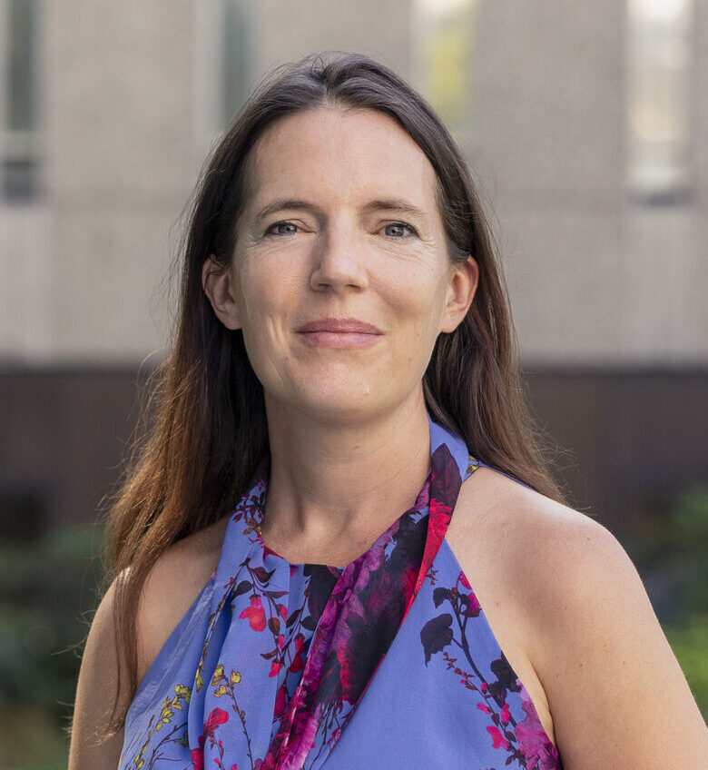
Molly Stevens
Professor of Biomedical Materials and Regenerative Medicine,
University of Oxford

Scott Delp
James H. Clark Professor
School of Engineering and Medicine
Stanford University
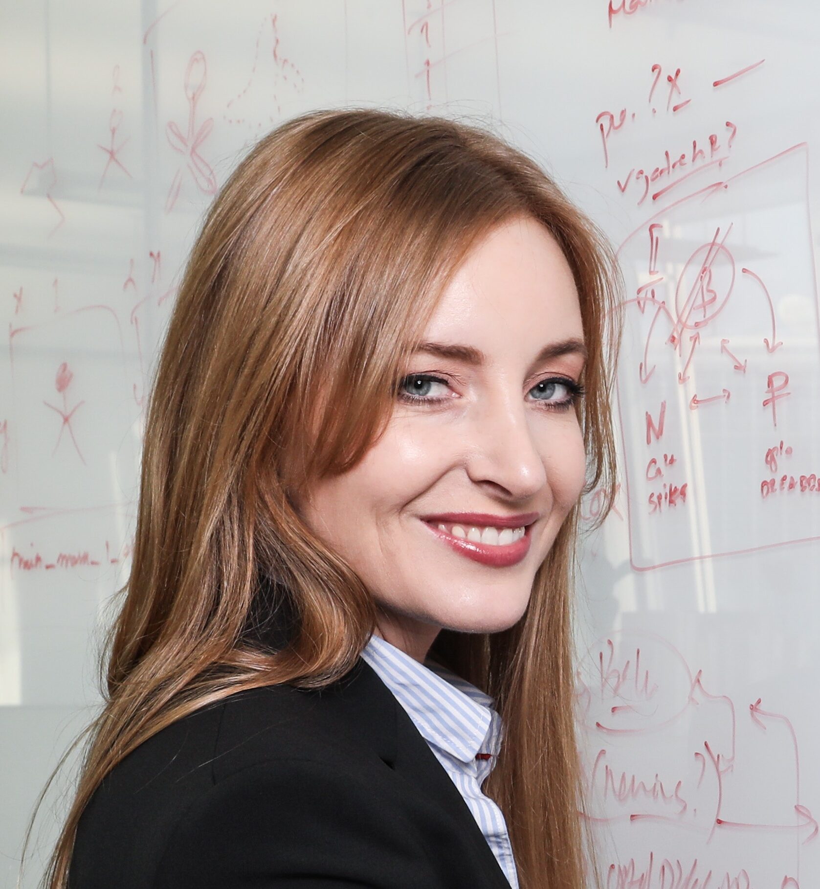
Mackenzie Mathis
Bertarelli Foundation Chair of Integrative Neuroscience Assistant Professor, Swiss Federal Institute of Technology
Wafa Skalli
Professor,
École Nationale Supérieure d’Arts et Métiers, Paris
Clinical Research in Spine Biomechanics

The spine is a key structure to withstand external loads, and particularly gravity in the erect position. It essential role is also to allow for motion while protecting the spine chord located in the medullar canal.
From a mechanical point of view, spine complex architecture has been widely investigated and modelled, taking into account the complex geometry and material properties of vertebrae and connecting soft tissues.
Despite these extensive efforts, clinical issues still represent major challenges for the Biomechanical engineer: spine disorders represent a huge human and societal burden, with associated pain impacting quality of life, fractures and loss of autonomy. When severe disorders require surgery, mechanical complications are not rare. Prevention is difficult because of the lack of understanding of the underlying mechanisms. In this context, clinically oriented research in spine biomechanics will be presented with two major aspects :
- Recent advances in subject specific spine modelling, considering not only the geometry and material properties of a given patient, but also subject specific loads, which require a global approach with head to pelvis and lowerlimb analysis in relation with musculo-skeletal issues.
- Translation from research to the service of patients and society. Academic research innovative models often require a tremendous effort (human and financial) to tackle issues such as extensive evaluation in real life (which may require drastic models evolution), proof of the medical service provided, ergonomy with regard to a routine clinical use, …. Some related research and development issues will be presented.
Wafa SKALLI is a Mechanical Engineer with a PhD in Biomechanics. She is currently Emeritus Professor at ENSAM. She was founder of the Institut de Biomécanique Humaine Georges Charpak, a place of convergence between clinicians and engineers, and holder of the BiomecAM ParisTech chair on subject specific musculoskeletal modeling. She is particularly involved in spine biomechanics and modelling, with a strong link to experimental and clinical approach. She is co-inventor of the innovative EOS low dose biplanar XRay system, developed in collaboration with Professors Jean Dubousset and Georges Charpak (Physics Nobel Prize 1992). She has been named as a free member (non-surgeon) of the National Academy of Surgery in France. Wafa Skalli is co-author of more than 350 referenced scientific publications and 13 patents. Her H index is 52. She is co-founder of the SKAIROS company, with the objective of bringing research advances to the service of patients and society.
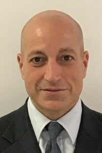
Fares S Haddad
Professor and Consultant Orthopaedic Surgeon,
University College Hospitals, London
Robotic Assisted Hip and Knee Arthroplasty: Are We Making Headway?
Conventional manual total hip arthroplasty (CO THA) and total knee arthroplasty (CO TKA) are well-established surgical procedures for the treatment of symptomatic end-stage arthritis. These procedures use preoperative two-dimensional radiographs for surgical planning and intraoperative handheld instruments to guide bone resections and component positioning. However, these manual procedures are associated with surgical errors in component positioning and have limited scope for assessing patient-specific joint biomechanics. Recent innovations in surgical technology have led to the development of robotic assisted total hip arthroplasty (RO THA) and robotic assisted total knee arthroplasty (RO TKA). These procedures use preoperative three-dimensional imaging to create patient-specific surgical plans and an intraoperative robotic device to execute these plans with high-levels of accuracy and reproducibility. In addition, RO THA uses dynamic preoperative assessments of spinopelvic kinematics to modify implant positioning, whilst RO TKA uses intraoperative assessments of knee biomechanics to fine-tune component positioning and restore native limb alignment. Existing studies have shown that RO THA is associated with improved accuracy in executing the planned horizontal and vertical centres of rotation, combined offset, cup inclination, cup version, and leg-length correction compared with CO THA. Clinical studies have shown RO TKA is associated with improved accuracy in templating component sizes, increased accuracy of component positioning, reduced opiate analgesia consumption, faster postoperative rehabilitation, shorter time to hospital discharge, less iatrogenic soft tissue injury, reduced systemic inflammatory response and improved Forgotten Joint scores at five years follow-up compared with CO TKA. For both RO THA and RO TKA, there are no learning curves for executing the planned component positioning and no additional risk of complications during the learning phases. Robotic planning software and intraoperative technology have been used to develop the coronal plane alignment of the knee (CPAK) and macroscopic soft tissue injury (MASTI) classifications, establish individualised “functional cup positioning” in THA and “functional alignment” in TKA, and assess the effects of controlled ligament releases and fixed flexion deformity on knee biomechanics.
Professor Fares S Haddad is professor of Orthopaedic and Sports Surgery and Divisional Clinical Director of Surgical Specialties at UCLH, and Director of the Institute of Sport, Exercise and Health (ISEH) at University College London, Medical Director of Orthopaedics, HCA UK and Editor in Chief, Bone and Joint Journal (formerly JBJS-Br).
Professor Haddad’s clinical and research endeavours have centred around hip and knee reconstruction. His interests include joint preservation after sports injuries, bearing surfaces, implant fixation, periprosthetic infection and outcomes assessment in hip, knee and revision surgery. His broader work also encompasses strategies to preserve and regain musculoskeletal health; he led the musculoskeletal team at the London Olympics 2012, was instrumental in setting up the National Centre for Sport & Exercise Medicine and has gained International Olympic Committee Centre of Excellence status at ISEH. He works with a number of elite sports and was the Chief Medical Officer for the NFL in the UK until 2022.
He was the gold medallist in the FRCS (Orth) exam and has gained a large number of prizes and prestigious academic awards. He has been an EFORT Travelling Fellow, British Hip Society Travelling Fellow and ABC Travelling Fellow in 2004. He was a Hunterian Professor in 2005. He is a member of the Hip Society, the Knee Society and of the International Hip Society (where he is currently President Elect).
He has presented and published widely on key aspects of hip, knee and sports surgery including over 700 peer reviewed publications. He leads a clinical research group with interests in joint preservation after injury, prosthetic design and performance and outcomes measurement after hip / knee injury, degeneration and surgery. He is Editor in Chief of the Bone and Joint Journal.
Alison Mardsen
Professor of Cardiovascular Diseases,
Stanford University, Stanford
Multi-physics Modeling in Pediatric Cardiology

Congenital heart disease affects 1 in 100 infants and is the leading cause of infant mortality in the US. Computational modeling is particularly valuable in this heterogeneous and high-risk population because of the need for personalized treatment planning. We will present recent work extending traditional hemodynamics simulations to include multiple physical processes and cardiac function in pediatric cardiology. In particular, we will discuss 1) melding constrained mixture models of vascular growth and remodeling with patient specific finite element simulations, and 2) multi-physics cardiac simulations incorporating electrophysiology, active contraction and fluid structure interaction. Novel algorithms for generating synthetic vascular networks for 3D bioprinting applications and simulating tissue perfusion will also be described. We will finally describe open-source software and data resources available via the SimVascular project and the Vascular Model Repository.
Alison Marsden is the Douglass M. and Nola Leishman Professor of Cardiovascular Disease in the Departments of Pediatrics, Bioengineering, and, by courtesy, Mechanical Engineering at Stanford University. She is a member of the Institute for Mathematical and Computational Engineering. From 2007-2015 she was a faculty member in Mechanical and Aerospace Engineering at UCSD. She graduated with a BSE degree in Mechanical Engineering from Princeton University in 1998, and a PhD in Mechanical Engineering from Stanford in 2005. She was a postdoctoral fellow at Stanford University in Bioengineering from 2005-07. She was the recipient of a Burroughs Wellcome Fund Career Award at the Scientific Interface in 2007, an NSF CAREER award in 2011. She was elected fellow of AIMBE and SIAM in 2018, the APS DFD in 2020, and BMES in 2021. She is the 2023 recipient of the Van C. Mow medal from the ASME Bioengineering Division. She has published over 170 journal articles and holds leadership roles in several scientific societies. Her research focuses on the development of numerical methods for cardiovascular biomechanics and application of engineering methods to impact patient care in cardiovascular surgery and congenital heart disease.

