CALL FOR ESB 2025 PRE-COURSES
If you are interested to organise a pre-course at ESB 2025 in Zurich, you can submit your proposal by 30th September 2024. Limited timeslots are available.
Fill in the application form and email it to info@esbiomech2025.org.
Fill in the application form and email it to info@esbiomech2025.org.
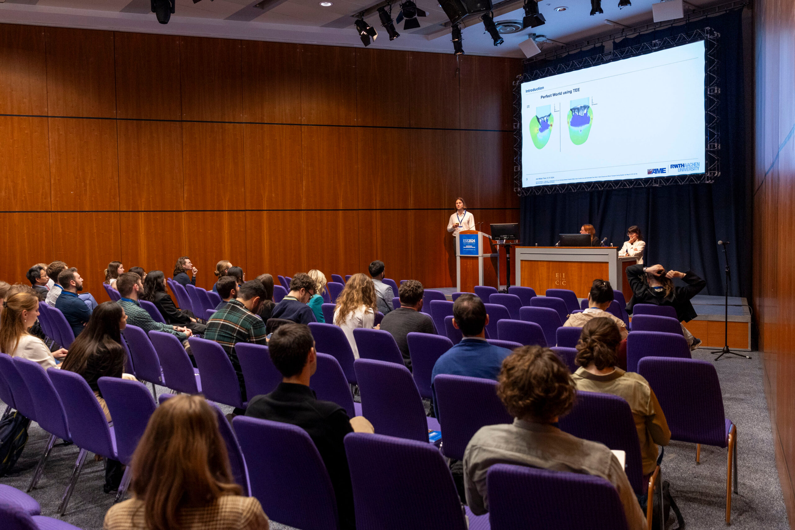
Learn by doing: expert-led ESB 2025 pre-courses await you!
ESB 2025 interactive pre-courses are mainly addressed to graduate students, post-docs, and young researchers.
Date: Sunday, 6 July 2025
Place: ETH Zürich Main Building
Registration for pre-courses is needed. The fee covers the organizational costs of the pre-course, lecturers do not profit from it.
Date: 6 July 2025, 13:00 – 15:00
Deep learning (DL) tools are increasingly integral to medical image analysis, particularly for shape representation. With rapid advancements in methodologies and network architectures, these tools leverage datasets to deliver cutting-edge results across a range of tasks. In this workshop, we will introduce participants to the fundamentals of shape representation using DL-based techniques through a mix of lectures and hands-on exercise. Additionally, we will cover Git version control to ensure that the developed DL-based models are traceable, reproducible, and aligned with the principles of open science.
Date: 6 July 2025, 13:00 – 15:00
This pre-course will cover a theoretical introduction, demos, and hands-on coding activities to automatically discover physics-based models from data. You should bring you own laptop and, if you like, your own data. We will provide benchmark data on brain, skin, arteries, and the heart, but are equally excited to help you analyse your own experiments. You will get the most out of this course if you familiarize yourself with the references and the code and prepare specific questions, but you are also welcome to attend if you are just curious about using artificial intelligence to explore biological systems.
Date: 6 July 2025, 13:00 – 15:00
Injuries happen in the blink of an eye but researchers in Injury Biomechanics believe most unintentional injuries are preventable. In this course the fundamental methods and knowledge used by injury biomechanics researchers will be presented in a hands-on interactive course.
Date: 6 July 2025, 15:30 – 17:30
BioDynaMo is a versatile, open-source simulation platform designed for modeling complex biological systems (https://www.biodynamo.org). It offers a flexible framework for building and simulating agent-based models, enabling researchers to explore a wide range of biological phenomena. Here, we will give an introduction into BioDynaMo and explain how the principles of agent-based modelling are supported. To this end, we will present several use cases to showcase the software capabilities. Afterwards, the team will run a hands-on tutorial session where a simple simulation is conducted. The course will conclude with an interactive conversation to address FAQs and any questions raised by the audience.
Date: 6 July 2025, 15:30 – 17:30
Biological hard and soft tissues are architectured or multiscale materials that hardly fulfil the definition of representative volume elements. Their behavior strongly depends on porosity that spans from nanometer to millimeter length scales as well as an anisotropic layout of their constituents. They are made of widely available materials, partly with inferior properties. Yet their composition and arrangement enable disparate material properties ranging from strengths of over 700 MPa to deformability of over 200%. Image-based tissue mechanics is an excellent tool-box to unlock such properties and study their dependence on aging and disease.
Date: 6 July 2025, 15:30-17:30
DeepLabCut is a deep learning-based python package for pose estimation that is open-source and free. It can be used on humans and other animals. It allows users to customize their models for specific keypoints (joints) of interest, or use a pretrained model. The workshop is tailored to new users of the software. It will include a short overview talk then hands-on practice with the code.
Background:
The analysis of anatomical structures in medical imaging is a cornerstone of research in biomechanics and biomedical engineering. Over the years, numerous methods have emerged, but the latest advancements are deeply rooted in data-driven deep learning (DL) approaches (e.g., fully connected networks for disease classificaton, convolutional neural networks for segmentation). This workshop will explore the versatility of DL-based methods for shape representation and analysis, demonstrating
their adaptability and scalability for a wide range of tasks. In addition, we will emphasize the critical role of Git-based version control, particularly through GitHub, to ensure that code is accessible, traceable, and reproducible.
Content and learning objectives:
Content: Basics of Shape Representation Using DL
LO: Participants will gain an understanding of the fundamental components of convolutional neural networks (CNNs), including layers, optimizers, and loss functions. They will also learn how to interpret training outcomes and adjust hyperparameters for improved performance.
Content: Advanced Frameworks for Shape Representation
LO: Participants will learn to analyze and interpret state-of-the-art network architectures and frameworks from recent literature. They will be equipped to replicate and adapt these models for their own research or applications.
Content: Git Version Control
LO: Participants will learn the essentials of Git version control using GitHub. By the end of the session, they will be able to create repositories, make commits, push and pull changes, and manage merges, ensuring a collaborative and efficient workflow.
Structure and duration (lecture, hands-on application …):
Shape Representation Methods (1.5 hours)
• Introduction to CNNs and Image Processing (Lecture)
An overview of CNNs and their role in medical image processing.
• Introduction to Advanced DL Models (Lecture)
Exploration of state-of-the-art DL models and their applications in shape representation.
• Implementing and Training an Advanced DL Model (Hands-on)
An interactive exercise to train a network based on a visual foundation model, allowing participants to gain practical experience.
Version Control with Git (0.5 hours)
• Introduction to GitHub and Version Control (Lecture)
An overview of Git, its importance in collaborative projects, and the fundamentals of GitHub for version control.
• Creating Repositories and Practicing Version Control (Hands-on)
An interactive exercise to help participants create their own repositories and practice key version control actions, such as commits, pushes and merges.
Potential attendees:
• PhD candidates and (early-career) researchers interested in applying deep learning to shape representation in their projects.
• Researchers heavily involved in (computational) modeling who face challenges with repeatability, version control, and managing code efficiently.
Requirements from the participants:
• Laptop with a GPU or access to Google Colab
• Software packages to download
• GitHub account
• Basic knowledge of linear algebra, statistics, calculus, and programming experience (Python)
Nazli Tümer is an Assistant Professor in the Department of Biomechanical Engineering at Delft University of Technology and a researcher in the Department of General Practice / Orthopedics & Sports Medicine at Erasmus Medical Center. Her research focuses on developmental skeletal biomechanics, utilizing both data-driven and mechanistic approaches.
Morteza Homayounfar is a researcher specializing in the development of DL models, including generative models, tailored for medical image processing. Currently pursuing a PhD at Erasmus MC and TU Delft, he is working on an advanced model that integrates shape, time, and density to study the hip growth.
Edwin Tay is a researcher with experience in the fields of multi-material 3D printing, finite element analysis, shape modelling, and DL. He holds an MSc in Biomedical Engineering from TU Delft and is currently pursuing a PhD there, focusing on spatiotemporal shape modelling for paediatric applications.
Background: Biological hard and soft tissues are architectured or multiscale materials that hardly fulfil the definition of representative volume elements. Their behavior strongly depends on porosity that spans from nanometer to millimeter length scales as well as an anisotropic layout of their constituents. They are made of widely available materials, partly with inferior properties. Yet their composition and arrangement enable disparate material properties ranging from strengths of over 700 MPa to deformability of over 200%. Image-based tissue mechanics is an excellent tool-box to unlock such properties and study their dependence on aging and disease.
Content and learning objectives: In this pre-course, participants will explore image-based tissue mechanics from hard to soft tissues in three parts:
1. from multi-scale images to properties,
2. from (time-elapsed) images to hard-tissue models, and
3. from images to soft-tissue models.
Learning aims are:
• [1 – analyses] understanding what information can be obtained for tissue mechanics by multiscale imaging data from hard and soft tissues (geometry, texture, microstructure, temporal and spatial evolutions, etc.),
• [2 – synthesis] understanding how these data can be used to feed computational or theoretical models for tissue mechanics,
• [3 – theorising] understanding differences and similarities between image-based hard and soft tissue mechanics.
Structure and duration: Three interactive sessions of 40 minutes including short breaks.
Potential attendees: Interdisciplinary graduate students involved in hard and soft tissue research.
Requirements from the participants:
Basic knowledge in applied mechanics, interest in image-based analyses and modelling, as well as a laptop.
Marta Peña Fernández obtained an MSc-level degree in industrial engineering from the University of Seville and a PhD in biomechanics from the University of Portsmouth. After postdoctoral positions at KTH and Heriot-Watt University, she is now Assistant Professor at Heriot-Watt University where she pursues her research on multiscale imaging and mechanics of mineralised tissues.
Hari Arora obtained his MEng and PhD in Mechanical Engineering from Imperial. After a Research Fellowship in Lung Mechanics at Imperial Bioengineering, he moved to Swansea University. He is now an Associate Professor leading the Biomedical Engineering Simulation and Testing Lab fusing experimental soft tissue biomechanics with advanced computational techniques.
Uwe Wolfram obtained an MSc-level degree in mechanical engineering from Chemnitz University of Technology and a PhD from the medical faculty of Ulm University. After positions at University of Bern and Heriot-Watt University he is now Professor at Clausthal University of Technology where he pursues his research on architectured mineralised tissues and digitalisation in materials science.
Background: BioDynaMo is a versatile, open-source simulation platform designed for modeling complex biological systems. It offers a flexible framework for building and simulating agent-based models, enabling researchers to explore a wide range of biological phenomena.
Content and learning objectives:
This pre-course will provide participants with a hands-on introduction to BioDynaMo by covering topics such as:
- Principles of agent-based modelling (ABM)
- Code structure and implementation
- Setup and usage of the software
- Basic simulation setup and configuration
- Several ABM demos
- Collaboration opportunities
By the end of the workshop, participants will have a solid understanding of ABM and BioDynaMo’s capabilities, and be able to apply it to their own research projects.
Structure and duration (lecture, hands-on application …):
Lectures following by a hands-on tutorial and an interactive discussion.
Potential attendees:
Anybody interested in agent-based modelling and applications for simulations in biological mechanics.
Requirements from the participants:
The participants should be able to run BioDynaMo. This can be done using an installed version on their laptop, or using a virtual machine, or in its browser-integrated form. Instructions for these are available here:
https://www.biodynamo.org/user-guide/from-installation-to-1st-steps
https://www.combynelab.com/home/research/biodynamo-introduction-materials
Dr Roman Bauer’s research focuses on the computational modelling and analysis of biological dynamics, in particular those of the brain. His highly interdisciplinary research involves modern computing approaches, biological expertise, innovative machine learning methods and IT- related collaboration. Dr Bauer is co-founder and spokesperson of the international BioDynaMo collaboration.
Dr Vasileios Vavourakis’ scientific expertise is in continuum mechanics, numerical methods, biomechanics, and in mathematical and computational modelling in engineering and bioengineering. His research interests extend across theoretical and applied science; more specifically in non-linear mechanics of biological tissues and biofluids, multiscale and multiphysics simulations, mathematical biology, and high-performance computing.
Background:
For more than 100 years, chemical, physical, and material scientists have proposed competing constitutive models to best characterize the behaviour of man-made and natural materials in response to mechanical loading. Now, computer science offers a universal solution: neural networks. Neural networks are powerful function approximators that learn constitutive relations from big data without any knowledge of the underlying physics. However, classical neural networks entirely ignore a century of research in constitutive modelling, violate thermodynamic considerations, and fail to predict the behaviour outside the training regime. In this pre-course, we introduce constitutive neural networks, a new family of neural networks that inherently satisfy common kinematic, thermodynamic, and physical constraints and, at the same time, constrain the design space of admissible functions to create robust approximators, even in the presence of sparse data. We show that this new class of network models is a generalization of the classical Neo-Hooke, Blatz Ko, Mooney Rivlin, Yeoh, Demiray, and Holzapfel models and that the network weights have a clear physical interpretation in the form of shear moduli, stiffness-like parameters, and exponential coefficients. To familiarize yourself with this new technology, you will implement your own constitutive neural network and train it with classical benchmark data, for example, from brain, skin, arteries, and the heart. You are welcome to bring your own data! You will see that your constitutive neural network autonomously selects the best constitutive model, parameters, and experiment to characterize your material. This new technology could have the potential to induce a paradigm shift in constitutive modelling, from user-defined model selection to automated model discovery. At the end of the course, we will share source codes, benchmark data, and documented examples.
Content and learning objectives:
In this short course, you will learn about automated model discovery; participate in a hands-on programming experience to implement and train your own constitutive neural networks; and receive a library of neural network notebooks to analyse and interpret classical benchmark data of living materials like human brain, skin, arteries, or the heart.
Structure and duration:
1. Brief history of constitutive modelling (20 minutes)
2. Introduction to constitutive neural networks (20 minutes)
3. Overview of mechanical testing (20 minutes)
Short break for discussion and Q&A
4. Automated model discovery for biological systems using your own Jupyter notebooks (60 minutes)
Potential attendees:
This pre-course is designed equally for Bachelor’s, Master’s, and PhD students, postdocs, and junior faculty who are interested in mechanical testing, analyzing data, modeling soft biological systems, and using machine learning and artificial intelligence to discover new knowledge from data, or in collaborating with our group and others to push the frontiers in automated model discovery.
Requirements from the participants:
Bring your own laptop if you want to participate in the hands-on automated model discovery part of the course. We will provide Jupyter notebooks, source codes, benchmark data, and documented examples, but you are welcome to bring your own data.
Ellen Kuhl is the Catherine Holman Johnson Director of Stanford Bio-X and the Walter B. Reinhold Professor in the School of Engineering. Her area of expertise is Living Matter Physics. She received the 2023 ERC Advanced Grant for her research on Automated Model Discovery. She is a passionate marathon runner and a multiple Ironman World Championship participant.
Mathias Peirlinck is Assistant Professor of BioMechanical Engineering at Delft University of Technology in the Netherlands. His area of expertise is soft tissue mechanics and in silico cardiovascular modeling. He received the 2023 Veni Talent Award for his research on data-driven and physics-based inference of myocardial tissue behavior.
Skyler St. Pierre is a Mechanical Engineering PhD Candidate in Professor Kuhl’s group. Currently, he conducts experimental tests on plant-based and animal meat and uses physics-based neural networks to model their constitutive behavior. He is funded by the NSF Graduate Research Fellowship and Stanford DARE Fellowship. He is also a triathlete.
Moritz Flaschel holds a PhD Degree in Computational Mechanics, which he acquired in Prof. Laura De Lorenzis’ group at ETH Zürich. In his doctoral thesis, he developed the method EUCLID, a method for automatically discovering interpretable material models from full-field data. Currently, he is a Postdoctoral Researcher in Prof. Ellen Kuhl’s group.
Background: Injury biomechanics is the subspeciality of biomechanics where practitioners design and improve safety equipment such as seat belts, airbags, ski bindings, helmets and vehicle seats and blast-protective footwear for current or legacy conflict regions. In this field an understanding of human biomechanics combined with an understanding of the injury tolerance and mechanical design are combined. The approaches and tools used in injury biomechanics research and practice are similar in some instances and very different in other instances compared to orthopaedic or sports biomechanics. In Injury Biomechanics we are often focused on impact events and human response that happen over a few milliseconds and generally less than 1 second. This necessitates impact test equipment, development of physical and computational surrogates that are biofidelic at impact load rates and high-speed data, video and x-ray collection. As the Injury Biomechanics field is small relative to other areas of biomechanics, the purpose of this pre-course is to introduce participants to the tools and approaches and some of the important findings from injury biomechanics in hopes of inspiring more students and staff to consider working in this field.
Content and learning objectives: Our learning objectives are for attendees to be able to:
- Describe several ways an injury biomechanics test suite would look different than a gait lab
- Describe the common tools used in injury biomechanics
- Describe the injury tolerance of three structures in the body (eg. head, thorax, extremities)
- Give two examples of novel injury prevention approaches that were presented in the course
- Name two injury mechanisms where a specific population is at higher risk and what is known of the reasons why.
Structure and duration:
- Tools used in Injury Biomechanics (30 minutes)
- Epidemiology
- Anthropomorphic Test Devices
- Cadaver Tissue Testing
- Computational Models
- Human Subjects
- Hands-on Exercise 1 (10 minutes)
- Part 2: Injury Tolerance (30 minutes)
- Head
- Spine and spinal cord
- Thorax
- Abdomen
- Extremities
- Hands-on Exercise 2 (10 minutes)
- Part 3: Injury Prevention (30 minutes)
- Helmets
- Seatbelts and airbags
- Body armour
- Hands-on Exercise 3 (10 minutes)
Potential attendees: All faculty and students attending ESB 2025
Requirements from the participants: None, the course will be pitched to a general biomechanics audience.
Dr. Spyros Masouros is Reader in Injury Biomechanics in the Department of Bioengineering, Associate Director in the Centre for Injuries Studies, and Director of the Injury & Reconstruction Test Suite at Imperial College London. Spyros’s research concerns the traumatic injury of the musculoskeletal system with a particular interest in the lower extremities, pelvis, and spinal column. The research combines clinical, experimental, and computational methods and technologies to determine mechanisms of injury, but also to assess current – and design new – treatment, protective, and rehabilitation strategies. Spyros is the President of the International Research Council on the Biomechanics of Injury (IRCOBI).
Prof. Dr. Kai-Uwe Schmitt trained as a paramedic, diplomas in mechanical engineering at University of Karlsruhe and Imperial College London, advanced studies in medical physics, doctoral degree and venia legendi for the field of Trauma Biomechanics at ETH Zürich. He manages the secretariat of IRCOBI.
Dr. Peter Cripton is a Professor in the School of Biomedical Engineering at the University of British Columbia. His research interests include impact biomechanics and injury prevention of the spine, spinal cord, brain and hip. Specific projects focus on improving bicycle helmet performance, developing improved mechanical surrogates for injury experiments (i.e. crash test dummy necks and physical models of the spinal cord), and using advanced MRI imaging techniques to better understand seat belt efficacy and sex differences in seat belt performance. Dr. Cripton is a member of the IRCOBI Council.
Background: DeepLabCut is a deep learning-based python package for pose estimation that is open-source and free. It can be used on humans and other animals. It allows users to customize their models for specific keypoints (joints) of interest, or use a pretrained model. The workshop is tailored to new users of the software. It will include a short overview talk then hands-on practice with the code.
Content and learning objectives: Learn about pose estimation in humans and other animals.
Structure and duration: There will be a lecture (45 min) followed by demos and hands-on time with the code.
Potential attendees: All faculty and students attending ESB 2025
Requirements from the participants: Python knowledge is a plus, but basic knowledge of coding is sufficient. Prior to the code participants can install DeepLabCut (deeplabcut.org), but it is not a strict requirement as they can use demo data in the cloud in the course.
The fee for the pre-course is covering the organizational costs of the pre-course, it is not going to DeepLabCut.
Prof. Mackenzie W. Mathis is the Bertarelli Foundation Chair of Integrative Neuroscience and an Assistant Professor at the Swiss Federal Institute of Technology, Lausanne (EPFL). Following the award of her PhD at Harvard University in 2017 with Prof. Naoshige Uchida, she was awarded the prestigious Rowland Fellowship at Harvard to start her independent laboratory (2017-2020). Before starting her group, she worked with Prof. Matthias Bethge at the University of Tübingen in the summer of 2017 with the support of the Women & the Brain Project ALS Fellowship. She is an ELLIS Scholar, Vallee Scholar, a former NSF Graduate Fellow, and her work has been featured in the news at Bloomberg BusinessWeek, Nature, and The Atlantic. She was awarded the FENS EJN Young Investigator Prize 2022, the Eric Kandel Young Neuroscientist Prize in 2023, The Robert Bing 2024 Prize, and the National Swiss Science Prize Latsis 2024.
Her lab works on mechanisms underlying adaptive behavior in intelligent systems. Specifically, the laboratory combines machine learning, computer vision, and experimental work in rodents with the combined goal of understanding the neural basis of adaptive motor control.

